In this piece PRISM researchers discuss how the use of artificial intelligence in pathology can open up new pathways towards the early diagnosis of cancer. The PRISM project has received funding from CRUK-STFC Early Detection Innovation Award to develop a data repository and associated AI tools to improve the diagnosis of malignant mesothelioma.
Pathologists can now use artificial intelligence (AI) algorithms to identify diseases more easily and quickly. Some of the most revolutionizing AI applications have been in medical imaging where researchers have, for example, used AI algorithms to detect retinopathy in medical images. Simultaneously, the improvement of technology resulting in, digital pathology, where glass slides are scanned and converted to digital images, allows the use of AI to improve diagnosis and prognostic processes in pathology. In cancer diagnosis and prevention these AI algorithms could identify abnormal cells that are either not visible to the human eye or that can only be recognised by pathologists after extensive training over many years. This could allow the routine identification of many more cancers at a pre-malignant stage increasing the number of cancers which can be prevented.
Why is pathology important for early cancer diagnosis?
Accurate and early diagnosis of diseases such as cancer is essential for optimal treatment. Patients with early stage disease have better life expectancy, compared to patients who may have a later diagnosis, when diseases such as cancer may have already spread to other organs or may have grown too big to cure. Especially, as new treatment modalities become available, accurate diagnosis will be increasingly essential for guiding therapy decisions and there will be an increasing need for alternative methods to accurately diagnose cancer and other diseases.
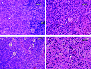
Currently, the actual diagnosis is still dependent on the correct interpretation of histological or tissue samples that are put on glass slides and examined by a pathologist under a normal microscope. Pathologists process and examine tissue biopsies and/or cells and make the diagnosis of diseases such as cancer. Using glass histopathology slides and microscopes, pathologists identify malignant or pre-malignant cells by recognising morphological features to help them classify any abnormalities according to specific morphological patterns and clinical features. Yet, even for pathologists with years of extensive systematic training and experience, the diagnosis of disease remains very subjective with differences in opinion being common between different pathologists.
Accurate histological diagnosis is often technically challenging because of the limited size of tissue samples, and difficulty in identifying “normal” cells from (pre)malignant cells.
Recent progress in Whole Slide digital Image scanners, which is the process of making digital photographs or digital images of the tissue samples on glass slides has created new and exciting opportunities for using these digital images of disease for better, faster and more accurate diagnosis. The large amounts of digital data that the digital images of tissue on the glass slides contain are also creating new opportunities for using AI.
What are AI, ML and DL?
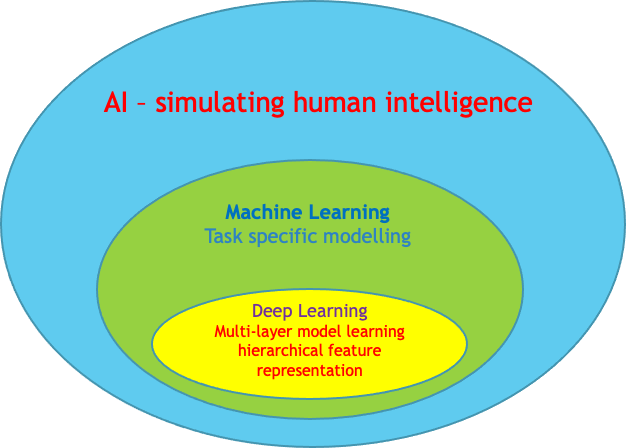
AI is the discipline concerned with the creation of intelligent software and machines. Intelligent software imitates human intelligence and can resolve informal tasks.
In the last few years, ML has developed faster due to the increased availability of data, and advances in computer hardware, e.g., GPU (Graphics Processing Unit). ML software extracts patterns from real-life data and develops its knowledge. Some of the most common ML tasks include classification, regression, transcription, machine translation, anomaly detection, denoising, density estimation and imputation of missing values.
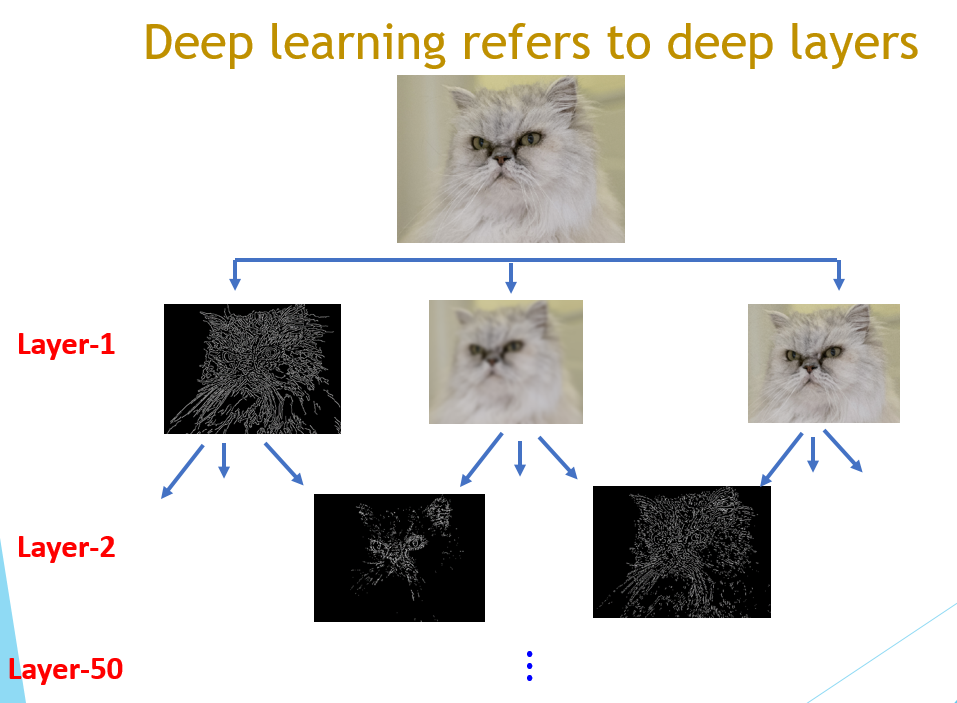
DL is a powerful ML technique and allows computers to understand our world in terms of taxonomy or hierarchy of concepts. For example, when identifying a face in an image, the software will build the primary concept “face” out of more simple concepts or features. Low-level features can be edges and dark spots; eyes, ears, and nose are considered mid-level features and the facial structure is a high-level feature. An example of such DL tools is the convolutional neural networks (CNN), also some of the first deep models that perform well in real-life situations.
Why can DL tools be more efficient than a human interpreter?
CNN is the means through which a computer can perform vision tasks. One of the essential operations of a CNN is the feature extraction process which also explains the efficiency of computers in image classification, segmentation and object recognition. By changing different parameters, we can choose what kind of features we extract from the image. A typical DL system applies more than 100 million parameters far beyond human comprehension.
One of the advantages of machines over human interpreters relies upon a computers’ ability to analyse an image at a pixel level that is not possible for the human eye. For example, in a 200.000 x 200.000 pixels image containing multiple types of cells, a deep learning algorithm could identify minimal anomalies. The information provided by the algorithm can be used by human interpreters in the decision-making process.
What are the roles of AI in histopathology?
ML algorithms are used for morphological and sub-visual (i.e., features that the human eye cannot see in a normal image of a cell or tissue sample) feature extraction and classification. This allows us to capture the three-dimensional histopathological properties displayed in whole slide imaging, which are essential for the next generation decision support systems in digital pathology. Especially sub-visual features are creating new pathways for diagnosing disease. The development of AI solutions for clinical image analysis and the identification of morphological features has also significantly advanced in recent years. The extraction and identification methods capture digital microscopic histopathological image properties at the pixel, object and even at higher semantic levels just the same way as a clinical expert would perform the tasks. These approaches form the essential core to the next generations of diagnostics for patients and form the basis of innovative AI-driven diagnostics.
There are several potential ways AI could be used in histopathology. Firstly, as an integral part of cancer screening and diagnosis, the demand for pathology increases every year, however, the workforce in the UK has not increased sufficiently to fill this demand. AI algorithms could be used to carry out certain diagnostic tasks, for example, identify and count certain types of cells, or predict genetic mutations and progression and outcome of cancers based on sub-visual signals, and even how certain disease will react to different therapies. Secondly, some cancers, like cervical cancers, have easily distinguishable premalignant stages (CIN1 to CIN3). This allows for prevention strategies where pre-malignancies can either be monitored or removed completely, preventing malignant cancer from developing. For other cancers, these premalignant cells can currently not be recognised by human pathologists, as they don’t have any known molecular markers or distinguishing features. Deep learning has the potential to identify distinguishing histological features which could be used as novel markers for pre-malignant cells.
The views expressed are those of the author. Posting of the blog does not signify that the Cancer Prevention Group endorse those views or opinions.
![]()